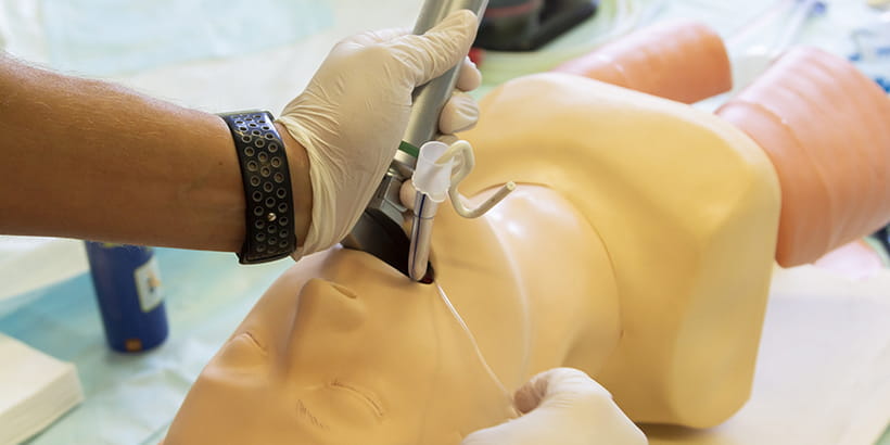
Endotracheal intubation can be successfully accomplished by video laryngoscopy (VL), flexible fiberoptic bronchoscopes, surgical techniques, and the classic direct laryngoscopy (DL), among others. DL has been the approach most commonly used by providers and has been utilized in operating rooms, emergency departments, ambulances, and intensive care units throughout the healthcare arena.
Successful laryngoscopy involves the distortion of the normal anatomic planes of the upper airway to produce a line of direct visualization from the operator’s eye to the larynx; this requires the creation of a new (nonanatomic) visual axis, through maximal alignment of the axes of the oral and pharyngeal cavities, and displacement of the tongue1. DL displaces pharyngeal soft tissues to create a direct line of vision from the mouth to the glottic opening2. Careful flexion of the neck and extension of the atlanto-occipital joint, or sniffing position, allows for proper alignment of the oral, pharyngeal, and tracheal axis3. Although atlanto-occipital extension cannot by itself allow direct laryngeal vision, it does provide anterior displacement of the mass of the tongue and brings up the alveolar ridge into an improved position relative to the tongue and larynx.1
The optimum positioning can be achieved by using pillows, blankets, or towels. The patient should be placed in the position that will be the most conducive for tracheal intubation (TI) on the first attempt; the first attempt should be the best attempt.
With the laryngoscope in the left hand, the patient’s mouth is opened with the first finger and thumb on the right hand using a “scissor” technique so the blade can be carefully placed in the right side of the mouth. The teeth need to be avoided as well as the lower lip. This can be accomplished by using the pinky finger on the left hand to move the lower lip down and out of the way of the blade. The tongue is swept to the left and up into the floor of the pharynx by the blade’s flange.3 The curved Macintosh blade is used to displace the epiglottis out of the line of sight by tensing on the glossoepiglottic ligament, whereas the straight Miller blade compresses the epiglottis against the base of the tongue.1 The Macintosh blade tenses the glossoepiglottic ligament by being placed in the vallecula. With either blade, the handle is raised up and away from the patient in a place perpendicular to the patient’s mandible to expose the vocal cords curved.2
A levering action should never be applied to the patient’s dentation due to the fact that such actions risk dental damage and diminish the quality of the laryngoscopic view.3 Extending either blade style too deeply can bring the tip of the blade to rest under the larynx itself so that forward pressure lifts the entire airway from view.1 A technique known as the Backwards-Upwards-Rightwards-Pressure (“BURP”) has been demonstrated to improve visualization of the vocal cords, and can be achieved by the clinician manipulating the larynx at the neck with his/her right hand during laryngoscopy.4
After successful DL, the properly sized endotracheal tube is placed between the vocal cords. The tracheal tube cuff should lie in the upper trachea, but beyond the larynx.2 The cuff’s pilot balloon is inflated to prevent an air leak. Over-inflation of the pilot balloon can cause damage to the tracheal mucosa. Pressures over 30 mmHg may inhibit capillary blood flow causing injury to the trachea.2
Confirmation of tracheal intubation is achieved by direct visualization of the endotracheal tube (ETT) passing through the vocal cords, end-tidal CO2, bilateral breath sounds heard over the chest in the midaxillary region, absence of breath sounds over the stomach, condensation with respirations in the ETT, and maintained or increased oxygen saturations. The ETT is secured at the appropriate depth, and radiographic confirmation is obtained if possible.
Tracheal intubation is a key step in airway management and can be accomplished in a variety of ways. It is up to the provider to recognize when this procedure needs to occur and to utilize the personnel best suited to accomplish the task safely and efficiently.
References
- Barash P, Barash PG, Cullen BF, Stoelting RK, Cahalan MK, Stock MC, Ortega R. Clinical Anesthesia Philadelphia. PA: Wolters Kluwer/Lippincott Williams & Wilkins.
- Butterworth JF, Mackey DC, Wasnick JD, Morgan GE, Mikhail MS, Morgan GE. Morgan & Mikhail’s Clinical Anesthesiology. New York: McGraw-Hill.
- Nagelhout JJ, Elisha S. Nurse Anesthesia. St. Louis, MO: Elsevier.
- Ulrich B, et al. The difficult intubation: The value of BURP and 3 predictive tests of difficult intubation. Anaesthetist. 1998;47(1):45-50.

